
Scientists explore how halophilic proteins survive high salt
 Proteins are a cell’s machinery. A cell’s surface is cluttered with protein receptors that lie waiting for environmental cues, which they then relay to interior proteins. These messanger proteins shuttle through the cell to yet other proteins poised to perform functions such as transforming cellular ingredients or altering gene expression. Cell life, and death, depends on proteins. And yet, proteins are often fragile. These long ribbons of chemical building blocks – the amino acids – must be kept in exactly the right arrangement of folds, coils, and loops. Most are held in place by no more than a transient network of positive and negative charges. Any deviation from this precarious assembly can be lethal, for the protein and potentially the cell. Changes in acidity, temperature, or salt concentration can be enough to tip the balance, but there are organisms, and their accompanying proteins, that survive such hostile environments, and scientists are eager to discover how.
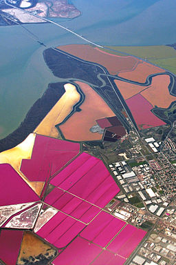
Extremophiles fascinate scientists because they defy expectations about what is necessary for, and what is prohibitive of, life. Halophiles are organisms that flourish where salt concentrations reach up to ten-fold what a typical cell can withstand. Some halophiles have evolved to maintain normal conditions inside the cell, but others survive by having a cellular interior just as salty as their surroundings. One such organism is Salinibacter ruber, which is a bacterium that was first found in salt crystallization ponds in Spain. This raises the question: how do the proteins withstand this assault of salt ions threatening to tear them apart?
To explore this question, a team of scientists in Grenoble recently compared two versions of a protein called malate dehydrogenase (MDH): one from S. ruber and one from Chloroflexus aurantiacus, another extreme bacterium that is adapted to high temperatures rather than high salt. The amino acid sequences of the two forms of MDH were 72% identical and their overall folds were likewise similar, but what interested the researchers most were the differences between the proteins. They found that S. ruber MDH had many more charged amino acids and, when they compared the structures using a technique called X-ray crystallography, they found that these adaptations occurred on the protein exterior.
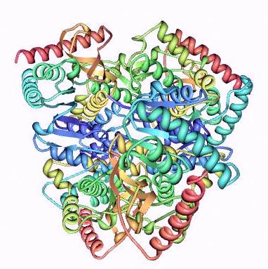
The team was particularly seeking to understand this interaction between the protein surface and the liquid immediately around the protein. The X-ray structure captured not only the folding of the protein, but also the organization of the neighboring water molecules. These findings show the first detailed comparison of the hundreds of water molecules that envelope a halophilic versus a non-halophilic protein. Like grease and water, nearly a third of the water molecules around the C. aurantiacus protein arranged themselves in pentagons to exclusively bind to each other, while meticulously avoiding contacts with the protein. S. ruber, on the other hand, showed no such polygons. The negatively charged amino acids allowed all of the nearby water molecules to bind directly to the protein surface, in a state of solvation.
While extremophilic proteins present a variety of potential applications for biotechnology, understanding their evolution also provides insight into the adaptive capacity of all life forms. From frozen glaciers to boiling sea depths to intolerable salt pools, life has found a way to exist, one molecule at a time. ~K.E.D.C.